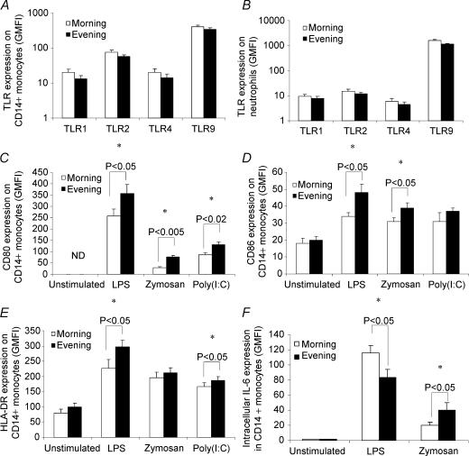
Figure 1. The effect of circadian rhythmicity on toll-like receptor (TLR) cell surface expression and activation.
Peripheral blood samples were obtained at 07.00 h and 17.00 h from eight healthy donors. A and B, samples were labelled with specific TLR monoclonal antibodies or isotype controls, and examined by flow cytometry. C–F, samples were incubated with media only (unstimulated), lipopolysaccharide (LPS; TLR4 ligand), zymosan (TLR2 and 6 ligand) or polyinosine-polycytidylic acid (poly(I:C); TLR3 ligand) for either 6 (D, E and F) or 24 h (C), and expression of costimulatory molecules and intracellular cytokines was examined by flow cytometry. All data represent means ± s.e.m.*Statistically significant difference as determined by paired-samples t test. ND, not detected.

