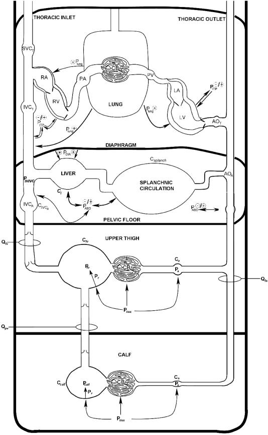
Figure 8. Schematic showing the sites of blood flow/velocity measurement and potential variables affecting the respiratory modulation of femoral and popliteal venous return.
For further explanation, please see Discussion and Appendix. LA, left atrium; LV left ventricle; AOT thoracic aorta; AOA, abdominal aorta; Csplanch, splanchnic vasculature compliance; Cl, liver compliance; Qfa, femoral arterial blood flow; Ca, arterial compliance; CB, capillary bed; Qfv, femoral venous blood flow; Cv, venous compliance; Ptiss, extravascular tissue pressure; Pr, recoil pressure due to elastic properties of vessel; QPV, popliteal venous blood flow (estimated by VPV in this investigation); IVCA, abdominal inferior vena cava, CIVCA, IVCA compliance; IVCT, thoracic inferior vena cava; SVC, superior vena cava; RA, right atrium; RV, right ventricle; PA, pulmonary artery; PV, pulmonary vein; PABD, abdominal pressure; PDIA, diaphragmatic pressure; PITP, intrathoracic pressure; Plung, lung surface pressure.

