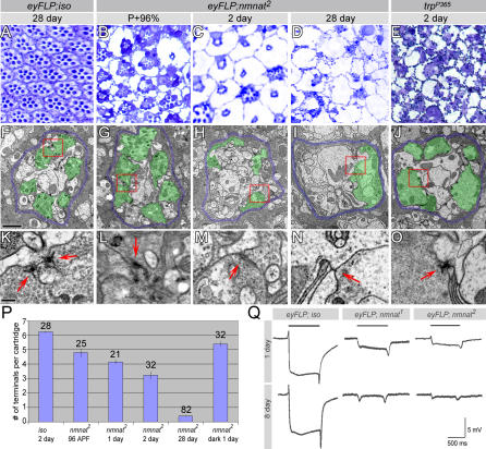
Figure 4. Loss of nmnat Causes Severe and Progressive Age-Dependent Degeneration.
(A–E) Retinal sections of control (iso), nmnat mutant eyes of different ages, and a trpP365 mutant eye. Reduced rhabdomeres and vacuoles are seen in P+96%, and both phenotypes become more severe with age. The nmnat mutant retina has a more severe phenotype than trpP365 in age-matched animals.
(F–J) TEM micrographs of lamina cartridges. Demarcating glia are colored blue and photoreceptor terminals green to accentuate the structures. In the mutant lamina, the number of structurally intact photoreceptor terminals gradually reduces with age. The trpP365 mutant lamina has a rather organized cartridge structure. The red boxes in (F–J) indicate the regions shown in (K–O), respectively. Scale bar in (F) for (F–J) indicates 1 μm.
(K–O) Individual synapses boxed in (F–J). In mutant photoreceptors, active zone structures gradually disintegrate with age (arrows); however, the morphology of the T-bars in trpP365 is well preserved. Scale bar in (K) for (K)–(O) indicates 200 nm.
(P) Quantification of the number of terminals per cartridge of control (iso) or mutant laminae at different ages. The photoreceptor terminals are recognized by the presence of capitate projections. In mutant laminae, the number of structurally intact photoreceptor terminals decrease with age, and dark rearing can delay the decline. The number of cartridges quantified is indicated above each graph.
(Q) ERG recordings of control (iso) and mutant photoreceptors at 1 d and 8 d of age. At 8 d, mutant photoreceptors have minimal responses to light.

