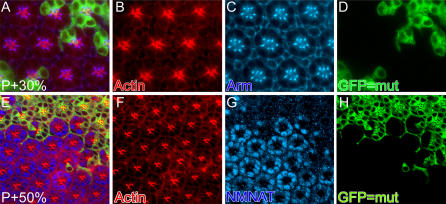
Figure 5. nmnat Mutant Photoreceptors Develop Normally.
MARCM analysis of pupal eye disc at 30 h after puparium formation ( P+30%) (A–D) and 50 h after puparium formation P+50% (E–H). GFP marks the mutant patch in (D) and (H). Anti-Actin antibody labels the rhabdomere structure in (B) and (F). Anti-Armadillo antibody labeling (Arm) marks the adherence junction in (C). Anti-NMNAT antibody shows labeling in wild-type cell bodies, but is dramatically reduced or absent in the mutant patch (G). In both developmental stages, there are no detectable structural differences between wild-type and mutant patches.

