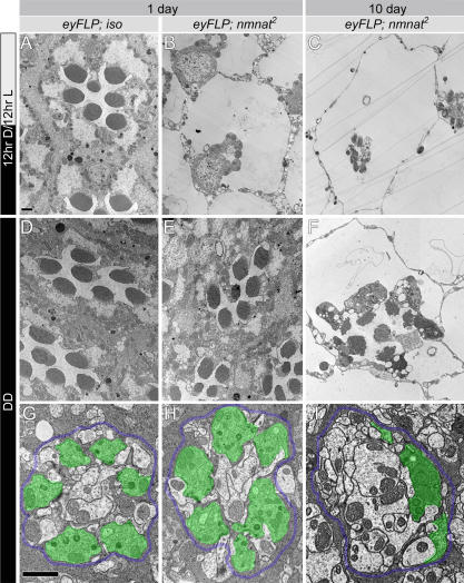
Figure 6. Light Enhances Neurodegeneration in nmnat Mutant Photoreceptors.
(A–C) TEM micrographs of control (iso) or nmnat mutant ommatidia at 1 d or 10 d of age kept in 12-hr light/dark cycle (12hr D/12hr L). Note the dramatic reduction of rhabodmere size at 1 d of age (B). This phenotype becomes more severe by day 10 (C). Genotypes and ages are marked on the top of each column.
(D–F) TEM micrographs of control or nmnat mutant ommatidia at 1 d or 10 d of age reared in constant darkness. Note the dramatic improvement at day 1 ([B] versus [E]) and day 10 ([C] versus [F]).
(G–I) TEM micrographs of cartridges containing control and nmnat mutant terminals at 1 d or 10 d of age reared in constant darkness. At 1 d of age, the mutant photoreceptor terminals are well organized, compared to the mutant photoreceptors of the same-aged flies raised in regular light/dark cycle (Figure 1H and 1I). Quantification of the number of terminals per cartridge is shown in Figure 4P. Dark rearing does not block synaptic degeneration, as at 10 d of age, the number of structurally intact photoreceptor terminals is reduced (I). Demarcating glia are colored blue and photoreceptor terminals green to emphasize the structures. Scale bars in (A) for (A–F) and in (G) for (G–I) indicate 1 μm.

