Figure 3.
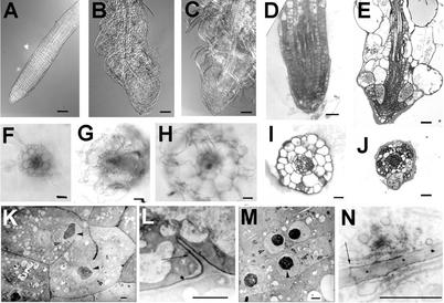
Root morphology of wild type (WT), ple, and hya. Whole-mount preparations of cleared WT (A), hya-3 (B), and ple-2 (C) root tips grown on nutrient agar medium using differential interference contrast microscopy. Median longitudinal and transverse sections of primary root tips of hya-3 (D and I) and ple-1 (E and J) resin embedded and stained with basic fuchsine/toluidine. In E and J, every tissue has aberrant cell wall stubs and multiple nuclei. Fresh transverse sections through the differentiation zone of a primary WT (F), hya-3 (G), and ple-2 (H) roots. The surface areas of hya roots are about 3 times and of ple-1 roots 3.5 to 4 times larger than WT. The three tissue layers epidermis, cortex and endodermis are clearly outlined but radially enlarged. H, In ple, the diarch symmetry of the vascular tissues is disrupted. K through M, TEM of ple-1 root cells. Note the darkly stained uneven nucleoli (arrowheads), the nuclear membrane (open arrows), and the incomplete cell walls. Longitudinal sections through cortex (K and N), endodermis (L), and vascular (M) cells. Although the newly developed cell wall is incomplete, suberin lamellas of the casparian stripe and plasmodesmata are incorporated (L and N, arrows). Bars = 50 μm in A through E, 25 μm in F through J, and 1 μm in K through N.

