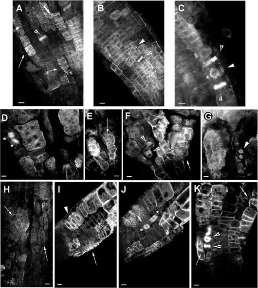
Figure 7.
Immunolocalization of microtubules in wild-type, ple-1, and hya-3 roots. A through C, Optical sections through the epidermis of wild-type roots showing cortical MTs (A; black arrow), phragmoplasts (B and C; white arrow), and the MTs of the PPB (arrowhead). The PPB is always accompanied with strong perinuclear MTs (C). D through G, Optical sections through the epidermis of ple-1 roots showing mitotic spindles (stars), dense accumulation of cytoplasmic MTs (D), cortical (E and F), and the perinuclear MTs of the PPB (G). Note the dark gray nuclei in multinucleated cells (D) and the cell wall stubs where MTs seems to nucleate (G). H through K, Optical sections through the epidermis and cortex of hya-3 roots showing slightly misoriented cortical (H, I, and K), perinuclear MTs and PPBs (I), mitotic spindles (J) of synchronized two nuclei containing cortical cells. K, Shows the misaligned phragmoplasts of a multinucleated epidermis cell. Bar = 10 μm.

