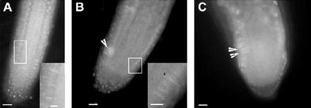
Figure 8.
Callose staining of cell plates in root meristems of wild type ple and hya. A, An epidermal cell file of wild type with callose in the cell plates. B and C, ple-2 and hya-1, respectively, show callose deposition in the cell plate of a synchronously dividing cells with multiple nuclei. Bars = 25 μm in A through C.

