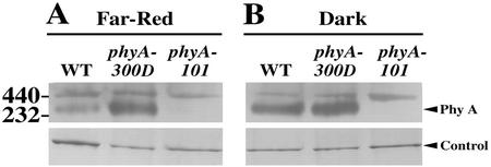
Figure 6.
Native gel analysis of phyA dimerization in FR and dark-grown wild-type and mutant seedlings. Extracts were separated on a 4% to 20% (w/v) gradient non-denaturing gel and probed with a polyclonal PHYA antibody. The phyA dimer is marked by a triangle on the right. Equal loading was ensured by visualizing a protein band staining intensity (marked by control). The positions of two mass markers are indicated on the left side. The phyA-101 is a null mutant control. A, FR light-grown seedlings (5 d); B, dark-grown seedlings (5 d).

