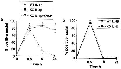
Figure 5.
IL-1-stimulated activation of NFκB is abnormal in iNOS KO BM cells. Nuclei were scored as positive or negative for NFκB staining in five fields from each well at ×20 magnification. (a) BM cells. (b) Osteoblasts. Differences between groups were analyzed by using Student's t test. *, P < 0.05; **, P < 0.01; ***, P < 0.001, all from WT at the same time point. Values shown are means ± SD for three replicates at each time point.

