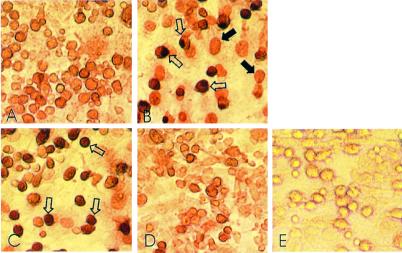
Figure 6.
Histochemical staining for NFκB nuclear translocation in cocultures. Filled arrows indicate staining of BM cell nuclei, and open arrows indicate staining of osteoblast nuclei. (A) iNOS KO coculture, unstimulated. (B) iNOS KO coculture, 30 min after stimulation with 10 units/ml IL-1. (C) WT coculture, 24 h after stimulation with 10 units/ml IL-1. (D) iNOS KO coculture, 24 h after stimulation with 10 units/ml IL-1. (E) Negative control showing absence of staining when cells were incubated with FCS instead of anti-p65.

