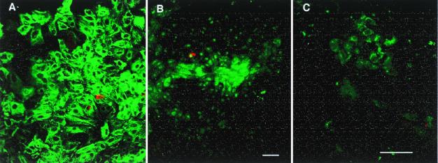
Figure 1.
(A) The adherent cells are primarily epithelial cells, immunostained for pan-cytokeratin (FITC green); staining for pan-cytokeratin and cytokeratin 19 were similar. Insulin-positive cells (Texas red) are scattered and infrequent. (B and C) Double staining of insulin (red) and transcription factor IPF-1 (FITC, green). Besides insulin-producing β cells, many duct cells express this transcription factor, both in the nucleus and in the cytoplasm. In B a number of cells express IPF-1 in the nucleus and/or cytoplasm without insulin staining; the field has the same density of cells as A. In addition, as in C, scattered clumps of cells had cytoplasmic IPF-1 staining with little nuclear staining and no insulin staining. Both A and B are 7-day cultured tissue of pancreas H99–12 pellet, whereas C is 7-day cultured tissue of pancreas H99–10 middle layer. (Magnification bars = 50 μm.)

