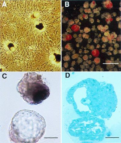
Figure 2.

(A) After ducts were overlaid with matrix, three-dimensional structures of ductal cysts with protruding buds of islet tissue (CHIBs) were observed rising from the monolayer lawn of cells. (B and C) There are variable numbers of dithizone-stained β cells in these harvested cycts/CHIBs; many of the structures are solely cysts whereas other have 50- to 150-μm islet buds. (D) The structure of budding islet cells from a cyst is seen in this toluidine blue 1-μm section. (Magnification bar = 500 μm in B, 100 μm in C, and 50 μm in D.)
