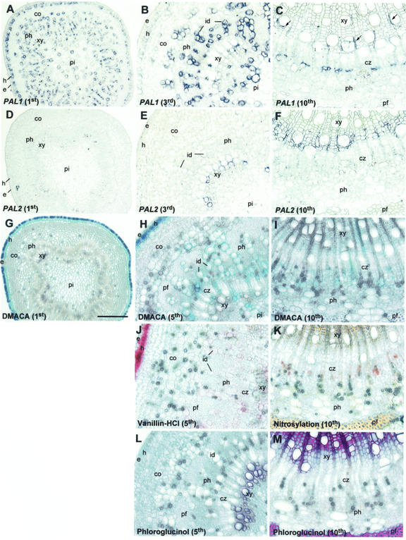
Figure 2.
In situ localization of PtPAL1 and PtPAL2 mRNAs and histochemical detection of CTs and lignin in aspen stem tissues. Transverse stem sections (10-μm thickness) were hybridized with digoxygenin (DIG)-labeled antisense PAL1 (A–C) or PAL2 (D–F) RNA probes and photographed in bright field. Transverse stem sections (75-μm thickness) were stained with dimethylaminocinnamaldehyde (DMACA; G–I), vanillin-HCl (J), or were nitroso-derivatized (K) for the detection of CTs, or were stained with phloroglucinol for the detection of lignin (L and M). Shown are first internode (A, D, and G), third internode (B and E), fifth internode (H, J, and L), and 10th internode (C, F, I, K, and M). Scale bar = 200 μm (A, D, and G) or 100 μm (all other panels). co, Cortex; cz, cambial zone; e, epidermis; h, hypodermis; id, idioblast, pf, phloem fibers; ph, phloem; pi, pith; xy, xylem.

