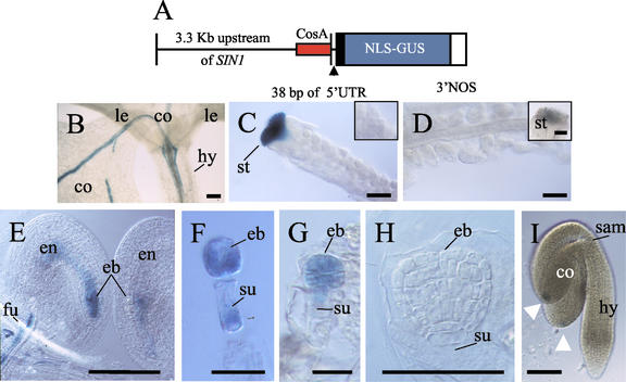
Figure 4.
Expression pattern of the SIN1/SUS1/CAF upstream genomic region, assayed by promoter GUS fusion. A, The SIN1/SUS1/CAF putative promoter construct pSP2, containing 3.3 kb of sequence upstream of the SIN1/SUS1/CAF gene (with 38 bp of the 5′-UTR), fused to the GUS reporter gene. The minimal promoter region from the cosmid CosA (which rescues the caf-1 mutant phenotype) is identified in red (Jacobsen et al., 1999). B, Two-leaf stage seedling (10 d post-germination) showing GUS activity in developing veins of the cotyledons and some expression in the hypocotyl. C, GUS expression is only detectable in the stigmatic tissue, and undetected in the initiating integuments at floral stage 11 (inset). D, Mature ovules at floral stage 13 show no detectable activity, and a reduction in GUS activity is seen in the stigma (inset). E and F, Post-fertilization ovules showing staining in the zygote and the endosperm. GUS activity is also present in the funiculus, which was observed throughout seed development. F, GUS expression in a dissected two-cell embryo, with activity in both the embryo and suspensor. G, GUS expression in octant embryo, but now absent from the suspensor. H, No detectable GUS activity in late globular or later embryonic stages (data not shown). I, Linear to cotyledon stage embryo showing GUS expression at the tip of the cotyledon (white arrowheads). co, Cotyledon; le, leaf; hy, hypocotyl; st, stigma; eb, embryo; en, endosperm; fu, funiculus; su, suspensor; sam, SAM. All bars = 100 microns, except F and G, which = 50 microns.

