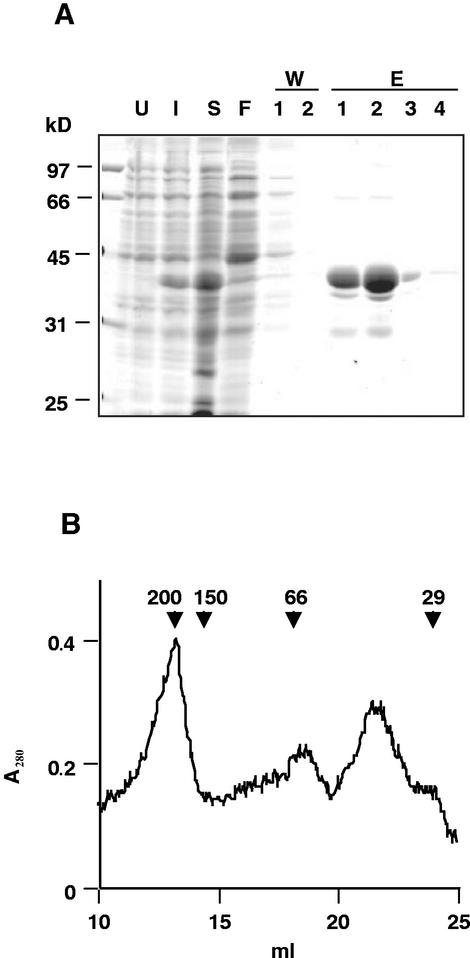
Figure 2.
Purification of DegP1. A, BL21-DE3 cells, transformed with pET-15b-DegP1 (U), were induced with 0.5 mm IPTG for 1 h (I). Cells were harvested, sonicated, and centrifuged to obtain a soluble fraction (S). This fraction was mixed with nickel-nitrilotriacetic acid (Ni-NTA) agarose and loaded onto a column, and the flow-through liquid was collected (F). The column was then washed twice (W1 and W2), and eluted four times (E1–E4) with 250 mm imidazole. Samples were resolved by SDS-PAGE on a 12% (w/v) acrylamide gel followed by Coomassie Blue staining. B, The pooled fractions E1–E4 were concentrated and loaded onto a Sephacryl S-200 column, and the A280 of the eluted protein was monitored. Relative migration of standard proteins and their molecular mass in kilodaltons is indicated by arrowheads.

