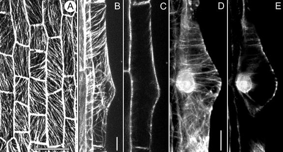
Figure 1.
CMTs in trichoblasts before bulge formation (A) and during bulge formation (B–E), visualized with a CLSM in scanning steps of 1 μm. A through C, GFP-MBD; D and E, Immunocytochemistry. A, CMTs are obliquely or longitudinally oriented to the long axis of the root. Bar = 50 μm. B, Full-stack projection of CMTs. CMTs loop through the tip of the bulge and are transversely or slightly helically oriented to the long axis of the root in the epidermal part. Bar = 10 μm. C, Projection of four median sections. There are no detectable EMTs in this developmental stage. D, Full-stack projection of CMTs; for explanation see B. Bar = 10 μm. E, Note position of the nucleus. For explanation see C.

