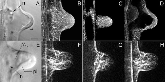
Figure 2.
MTs during transition from a bulge into a growing root hair, visualized with a CLSM in scanning steps of 1 μm (B–D and F–H). B through D, Immunocytochemistry; F through H, GFP-MBD. In this developmental stage, polar growth is initiated. A, Corresponding bright-field image to B through D; the bracket indicates a thin layer of cytoplasm at the tip. B, Full-stack projection of MTs. C, Projection of three peripheral sections showing CMTs. A few CMTs still reach the very tip and are net-axially oriented below the tip region. D, Projection of four median sections; EMTs start to appear at this stage of hair development. E, Bright-field image of a hair at a somewhat later stage than shown in A. The cytoplasmic layer in the tip region has increased in length. F, Full-stack projection of MTs. G, Projection of three peripheral sections. Single CMTs still reach the very tip. H, Projection of four median sections showing EMTs. There is a concomitant increase in the length of the subapical cytoplasmic dense region and the density of EMTs. A through D show an earlier stage than E through H. Magnification is the same in all images. Bar = 20 μm. n, Nucleus; v, vacuole; pl, cytoplasmic layer.

