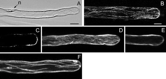
Figure 5.
MTs in full-grown root hairs visualized with a CLSM in scanning steps of 1 μm. B and C, Immunocytochemistry; D through F, GFP-MBD. A, Bright-field image of a living hair. Note position of the nucleus (n). Bar = 10 μm. B, Full-stack projection of CMTs. CMTs, the only remaining population of MTs in this developmental stage, are longitudinally oriented; they converge at the very tip. Bar = 10 μm (B–F). C, Projection of two median sections. Full-grown hairs have no EMTs. D, Full-stack projection of CMTs. The hair has stopped growth recently and the density of CMTs, which are net-axially oriented, is similar to previous developmental stages. E, Projection of two median sections; for explanation see C. F, Full-stack projection of CMTs in a hair of a later stage than shown in D. The density of CMTs is lower than in hairs that have just terminated growth.

