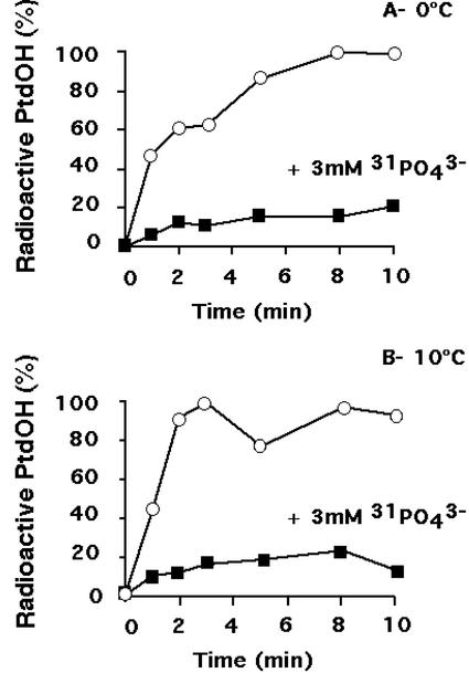
Figure 5.
Relative level of radioactive PtdOH formed in absence (white symbols) or presence (black symbols) of 3 mm nonradioactive PO43− during an exposure at 0°C (A) or 10°C (B). Phospholipids were labeled in presence of 53 MBq L−1 [33P]-PO43− for 105 min at 22°C. Nonradioactive PO43−, if required, was then added. Fifteen minutes later, cells were transferred at 0°C or 10°C. Lipids were extracted at different times after the beginning of the cold treatment, and were separated by TLC using an acidic solvent system. PtdOH content was quantified as a fraction of total radiolabeled phospholipids. PtdOH formed was normalized to the maximum amount detected in the absence of nonradioactive phosphate. A typical experiment for each condition is shown.

