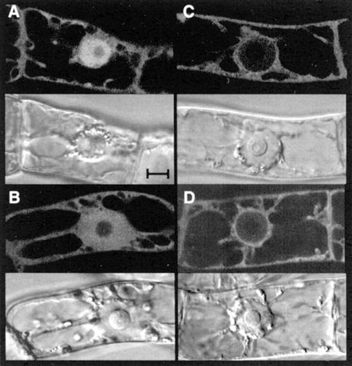
Figure 7.
Subcellular localization of UBC19 using GFP as life marker in tobacco BY2 cells. Each fluorescent focal plane (top panels) is shown with its corresponding transmitted light reference image viewed by differential interference contrast (bottom panels). Localization of GFP alone (A), UBC19-GFP (B), GUS-GFP (C), and UBC19-GUS-GFP (D) in interphase BY2 cells after 24 h of Dex induction. Bar = 10 μm.

