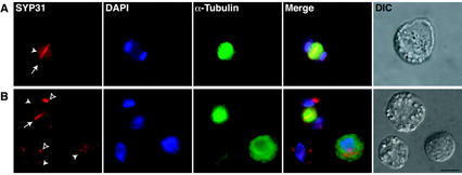
Figure 6.
Localization of SYP31 in dividing Arabidopsis cells. Arabidopsis cells were double immunolabeled with anti-α-tubulin antibodies (green) and affinity-purified anti-SYP31 antibodies (red) and DAPI. Merged images were generated electronically from the three preceding pseudocolored images and are shown in the indicated panels (merged). Differential interphase contrast images of the cells analyzed are presented (DIC). White arrows indicate the location of the cell plate. White arrowheads indicate the position of large undefined subcellular structures. Unfilled arrowheads indicate the position of small cytoplasmic punctate structures. Bar = 50 μm.

