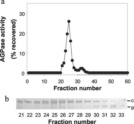
Figure 6.
Separation of isoforms of AGPase by ion-exchange chromatography. Wheat endosperms were extracted and the homogenate applied to a Q-Sepharose column as described in “Materials and Methods.” a, The activity of AGPase in each fraction is expressed as a percentage of the total AGPase activity recovered in all fractions from the column. b, Samples of fractions 21 through 33 (containing AGPase activity) were subjected to SDS-PAGE, blotted onto nitrocellulose, and probed with Bt2 antiserum at a dilution of 1/5,000. c, Cytosolic SSU; P, plastidial SSU.

