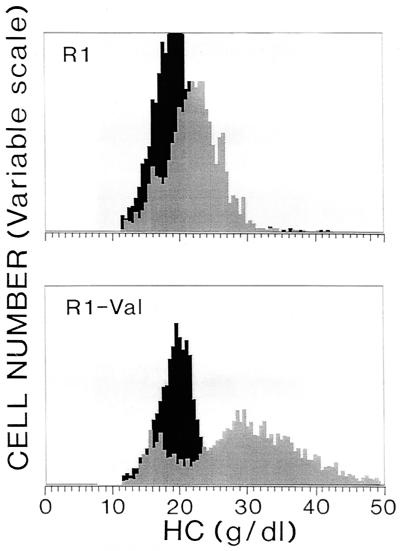
Figure 3.
Effects of K+-permeabilization with valinomycin on the HC distributions of the reticulocyte and nonreticulocyte components of the lightest, R1, fraction of SS RBCs. The treatment was the same as in Fig. 2, except that the samples of RBC suspension were diluted in buffer D containing RNA stain for the reticulocytes and incubated 15 min before adding the sphering agent. In the histograms, the reticulocytes are shown in gray and nonreticulocyte RBCs in black.

