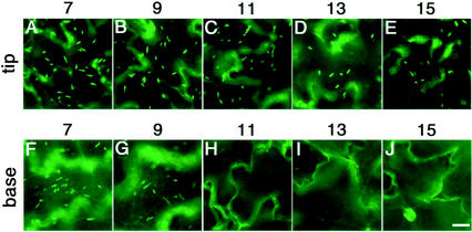
Figure 2.
The number of ER bodies in the epidermal cells of the cotyledons decreased during senescence. The tip part (A–E) and the basal part (F–J) of the cotyledons were inspected with a fluorescence microscope. The number of days after germination is indicated at the top. Disappearance of ER bodies started from the basal part of the cotyledons. The tip part (A–E) had ER bodies even at 15 d after germination, whereas the basal part (F–J) lost them in the 11-d-old cotyledons. Bars = 20 μm.

