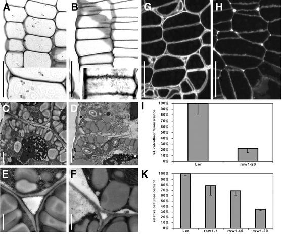
Figure 5.
Cell wall structure and composition in wild-type and rsw1-20 mutant embryos. A and B, Periodic acid-Schiff's stained longitudinal sections in the hypocotyl of bent-cotyledon stage embryos. Note the presence of intercellular spaces in the wild type (A), which are absent in the mutant (B), and the uneven, granular appearance of mutant cell walls (inset). C thorough F, Epidermal cell walls from hypocotyls of bent-cotyledon stage embryos of wild type (C and E) and rsw1-20 (D and F). Note that mutant cell walls are thinner and that the middle lamella is usually not discernible. Intercellular spaces in the wild type (E) are usually filled with wall material in rsw1-20 mutants (F). G and H, Calcofluor stained cross sections in the hypocotyl of bent-cotyledon stage wild-type (G) and rsw1-20 mutant (H) embryos. Overall reduced staining in mutant walls indicates reduced β-glucan content. Note the uneven staining intensity in mutant cell walls. I, Densitometric quantification of cell wall β-glucan content measured on digital images from calcofluor-stained hypocotyl cells of bent-cotyledon stage embryos. K, Spectrophotometrical determination of β-glucan content in wall preparations of wild-type and mutant seedlings after germination and growth for 7 d at 31°C. A and B, Bright-field optics images; C through F, transmission electron micrographs; and G and H, epifluorescence optics. Scale bars = 20 μm in A, B, G, and H; insets, 2× magnification; 1 μm in C and D; and 500 nm in E and F.

