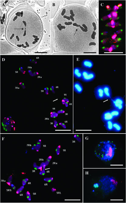
Figure 1.—
(A) PMC of sy10 at metaphase I showing a bivalent with a single, distal chiasma (arrow) and 12 univalents. (B) PMC of sy10 at metaphase I showing a “sticky” complex of three univalents (arrow). (C) Projection of an optical stack through a typical PMC at metaphase I of Sy10 wild type. The chromosomes are probed with pSc200 (red), pSc250 (blue), 25S rDNA (yellow), and CCS1 (green). (D) Projection of an optical stack through a PMC at metaphase I of sy10 hybridized in situ with the same probes as in C. All chromosomes are identified. The two chromosomes 4R are connected by a chromatin bridge (arrow) and constitute the only rod bivalent in this cell. (E) PMC in D stained with DAPI, which reveals the chromatin bridge between the two 4R chromosomes. (F) Similar cell to that in D, but showing a single ring bivalent (arrow) between two nonhomologous chromosomes. (G) Typical PMC at premeiotic interphase hybridized in situ with centromeric (red) and telomeric (green) probes and counterstained with DAPI. (H) Similar cell to that in G but at leptotene, showing a tight telomeric cluster (bouquet) colocalizing with DAPI-positive telomeric heterochromatin. Bars, 10 μm.

