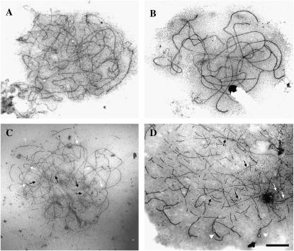
Figure 2.—
Electron micrographs of SCs of Sy10 wild type, showing the normal progression of synapsis from early (A) to late (B) zygotene. (C) Surface-spread PMC of the sy10 mutant at a stage equivalent to zygotene, showing widespread asynapsis and some short stretches of SC (solid arrows). Switches of pairing partners indicating indiscriminate synapsis are delimited by open arrows. (D) Late diplotene in the sy10 mutant showing fragments of axial cores and remnants of SCs (solid and open arrows, respectively) and foldbacks (arrowheads). Bar, 5 μm.

