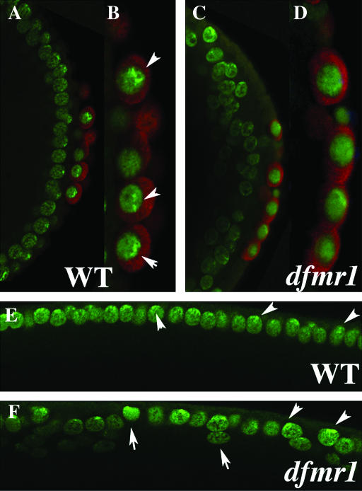
Figure 7.—
Localization of HP1 in dfmr13 embryos. (A, B, and E) Wild-type and (B, D, and F) dfmr1 syncytial blastoderm-stage embryos were labeled with HP1 (imaged in green) and Vasa antibody (imaged in red). (A–D) Merged images. (E and F) The HP1-specific staining. As described in the text, HP1 is localized in a punctate pattern in wild-type pole cells (see arrowheads) while its localization is much more diffuse in dfmr13 pole cells. In the case of somatic nuclei, HP1 is concentrated in a punctate pattern at the apical surface of wild-type nuclei. The distribution and level of HP1 in dfmr13 embryos varies from one nuclei to another. Also note that unlike wild-type nuclei, dfmr13 nuclei are not always elongated along the apical–basal axis and are often displaced from the surface (see arrows and arrowheads in F and somatic nuclei in C).

