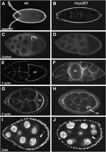
Figure 1.—
Phenotypes displayed by mus301 mutants. (A) Wild-type egg shell. (B) Fully ventralized egg shell. (C) Wild-type S9 egg chamber showing Gurken protein localization. (D) Mutant egg chamber stained and imaged under the same conditions as in C. No Gurken can be detected. (E and G) Wild-type egg chambers stained with rhodamine–phalloidin to visualize the morphology of cells. Both oocytes (oo) are located at the posterior of the egg chamber. (F and H) Mutant egg chambers labeled with rhodamine–phalloidin showing a misplaced oocyte (F) and a small, misplaced oocyte (H). (I) Wild-type follicle with the oocyte's chromatin condensed into a karyosome (open arrowhead). (J) Mutant karyosome. (B and D) mus301094/Df(3L)66C-G28. (F) mus301422/mus301660. (H) mus301D1/mus301D2. (J) mus301660/Df(3L)66C-G28.

