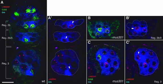
Figure 5.—
A role for mus301 in recombinational DSB repair. Wild-type (A) and mutant (B and C) germaria showing the distribution of γ-HIS2AV (red), C(3)G (green), and Orb (blue) proteins. (A) One cyst each from region 2b, region 2b/3, and region 3 is shown (see Figure 6 for a schematic of these germarial stages). γ-HIS2AV foci are abundant in region 2b oocytes, but they are lost in successive stages and are barely detectable in region 3 oocytes. (B and C) γ-HIS2AV foci are visible in region 2b/3 and region 3 mutant oocytes. The sibling nurse cells also possess increased γ-HIS2AV staining compared to wild type. All images are projections of several confocal sections. Dashed lines delineate individual cysts. Arrowheads point toward oocytes. (B and C) mus301094/Df(3L)66C-G28. Bar, 10 μm.

