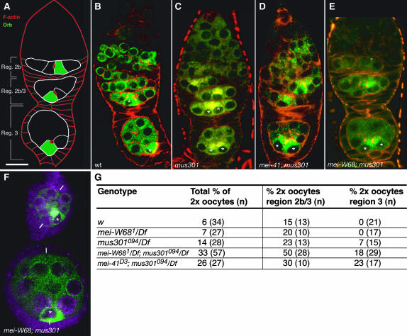
Figure 6.—
mei-W68 does not rescue the two-oocyte phenotype of mus301 mutant cysts. (A) Scheme of a wild-type germarium to show the arrangement of region 2b, region 2b/3, and region 3 cysts. (B–E) Germaria double stained to visualize filamentous actin and Orb protein. (B) Wild-type germarium showing Orb accumulated in a single cell in region 2b and region 3 cysts. (C) Mutant germarium carrying region 2b and region 3 cysts, each containing two cells that accumulate Orb. (D and E) Double-mutant germaria showing region 3 cysts with two cells containing high levels of Orb protein. (F) Double-mutant egg chambers stained with anti-Orb to show a misplaced oocyte. The white bars mark the anterior–posterior axes of the follicles. (G) Germaria of different genetic combinations were analyzed and the number of region 2b/3 and region 3 cysts containing two cells with increased levels of the oocyte marker Orb and each possessing four4 ring -canals (as visualized with rRhodamine–-pPhalloidin) were counted as “2× oocyte” cysts. (B) Wild type. (C) mus301094/Df(3L)66C-G28. (D) mei-41D3/Df(2R)LL5; mus301094/Df(3L)66C-G28. (E and F) mei-W681; mus301094/Df(3L)66C-G28. Asterisks label the cells with highest Orb contents. Bar, 10 μm.

