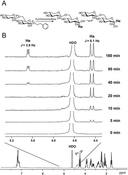
Figure 7.
Time course of hydrolysis of 2-phenylethyl β-primeveroside catalyzed by the tea leaf β-primeverosidase, followed by 1H-NMR spectroscopy. A, Postulated retaining hydrolysis of 2-phenylethyl β-primeveroside by the tea enzyme. B, Spectra recorded 0, 5, 10, 20, 40, 90, and 180 min after addition of the enzyme. Ha and He indicate the resonances of H-1 of α- and β-d-Glc, respectively. The full-scan 1H NMR spectrum of the substrate (2-phenylethyl β-primeveroside) is shown at the bottom.

