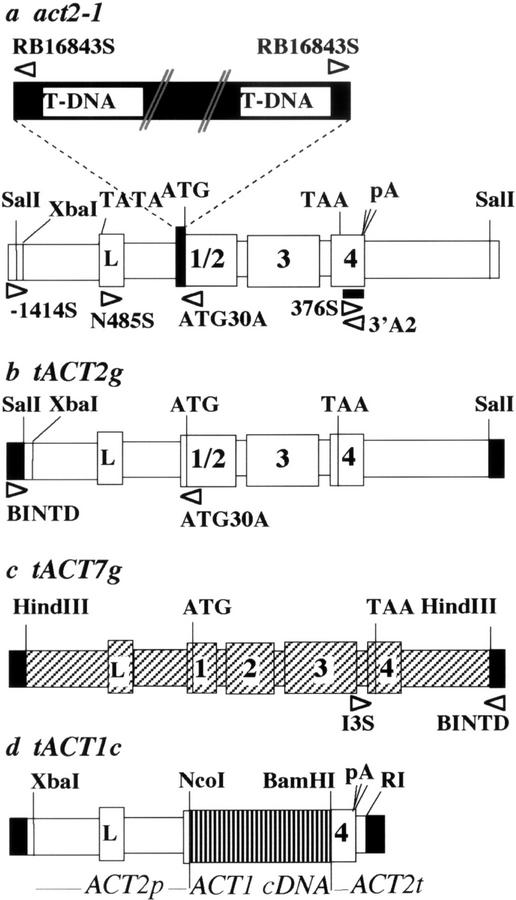
Figure 1.
Map of act2-1 mutant allele and complementing transgenes. a, The act2-1 allele contains a T-DNA insertion (black) separating most of the ACT2 5′-UTR (white) from the body of the ACT2 actin coding sequence (white rectangles, with exons drawn larger than introns or flanking sequence). The T-DNA insertion event deleted the acceptor splice junction including 12 nucleotides of the leader (5′-UTR) intron and four noncoding nucleotides of exon one-half of the act2-1 gene, leaving just five nucleotides between the T-DNA and the act2-1 start codon. Because the insertion is flanked on either side by a right border sequence, the allele contains two or more T-DNAs in tandem at this site. b, The genomic ACT2 transgene (tACT2g) used to complement the act2-1 mutant was contained on a 4-kb SalI fragment (white) flanked by T-DNA sequences (black). c, The genomic actin ACT7 transgene (tACT7g) used to complement the act2-1 mutant was contained on a 4-kb HindIII fragment (diagonal lined rectangles). d, The cDNA transgene, tACT1c, used to complement the act2-1 mutant, contained the actin ACT1 protein coding sequence (vertical lined rectangles), under control of the ACT2 (white) promoter (ACT2p) and terminator (ACT2t) sequences (Kandasamy et al., 2002). The white arrowheads refer to positions and orientations of PCR primers and the thick horizontal black bar (a) refers to the location of the 3′-UTR DNA probe used for northern analysis.

