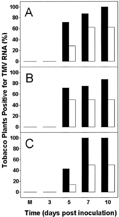
Figure 4.
Summary of northern results establishing the presence or absence of TMV coat protein RNA in leaf tissue from wild-type (A), AS8 (B), and S24 (C) plants. Data represent the percentage of plants with detectable TMV coat protein RNA. Plants were grown in the absence (black bars) or presence (white bars) of SA and were inoculated with TMV, as explained in the legend to Figure 3. Total RNA was isolated from leaves 2 and 3 above the inoculated leaf, and 5 μg of RNA was separated on agarose gels containing formaldehyde and used for northern analysis with a radiolabeled DNA probe recognizing TMV coat protein RNA. This allowed plants to be readily scored for the presence or absence of TMV coat protein RNA. The M refers to a plant that was mock inoculated, in which case RNA was isolated at 10 DPI. Each bar represents data obtained from eight separate plants, each from a separate experiment.

