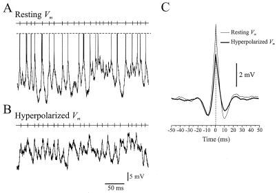
Figure 3.
Membrane hyperpolarization decreases the amplitude and rate of rise of transient depolarizing potentials evoked by visual stimulation. (A) The Vm of a layer II/III regular spiking pyramidal neuron (simple cell) recorded during a single presentation of a drifting grating. The action potentials have been truncated as indicated by the dashed lines. (B) The response of the same cell to a single presentation of the stimulus, while it was hyperpolarized with a steady negative current to block spike discharges (−2.0 nA). The upper trace in both A and B indicates the time of occurrence of each local maximum in the median-filtered Vm used to compute the cycle-triggered-average. (C) Cycle-triggered averaging reveals that the transient depolarizations evoked by visual stimulation were greater in amplitude and rate of rise during control conditions (6.7 mV, 1.34 mV/ms) than during hyperpolarization (4.5 mV, 1.12 mV/ms).

