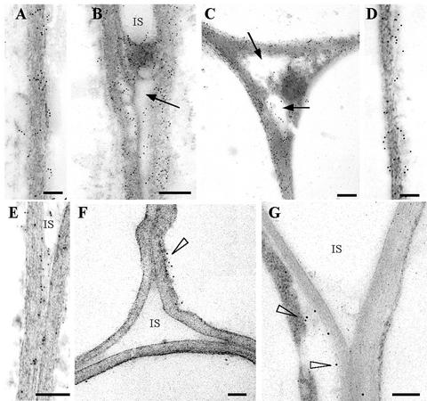
Figure 6.
Immunogold labeling of leaf parenchyma cell walls 4 (A, B, and E) and 7 (C and D) d postinfiltration with A. tumefaciens carrying pExp::PG1ΔSP (A–D) or the mutant pD202N/D203N (E). Labeling was achieved with antiserum against CLPG1 and gold-conjugated goat antiserum to rabbit IgG. Gold particles were found within the cell walls (A and B) and in intercellular spaces (C and D) at nearly identical levels at 4 and 7 dpi. Note degradation of pectic material in intercellular spaces (IS; arrows). Gold particles were also observed within the cell walls and intercellular spaces 4 dpi with A. tumefaciens carrying the mutant pD202N/D203N (E), without detectable pectin degradation. A few gold particles were observed in leaves agro-infiltrated with the empty vector (cell wall-cytoplasm interface, arrowhead, F), and in sections treated with the secondary antibody alone (arrowheads, G). Sections were contrasted with uranyl acetate. Scale bar = 0.3 μm.

