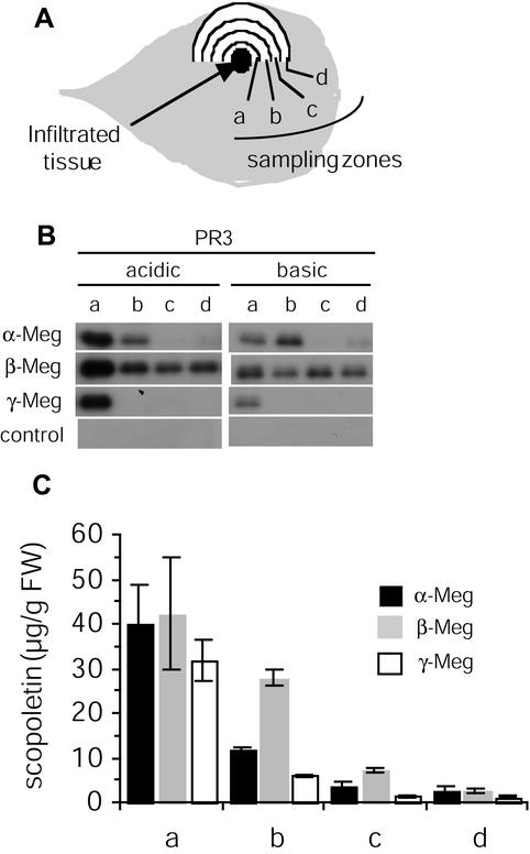
Figure 6.
Acidic and basic PR3 protein expression and scopoletin accumulation in tissue exhibiting LAR after treatment with α-, β-, or γ-megaspermin. Tobacco leaves were infiltrated with 50 nm α-, β-, or γ-megaspermin or water. Tissues in the vicinity of the infiltrated tissues were collected 24 h after treatments and analyzed. A, Diagram showing the sampling zones. Each zone is 5 mm wide. B, Acidic and basic PR3 protein immunodetection after western blotting. C, Total (free + conjugated forms) scopoletin accumulation quantified by HPLC. No scopoletin was detected in control plants. Results have been expressed as the mean and sd calculated from two independent experiments.

