Figure 2.
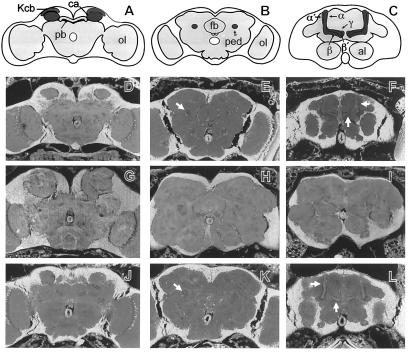
Rescue of the brain anatomy phenotype of mud by a genomic fragment. (A-C) Schematic frontal sections of wild-type Drosophila heads at the level of the calyces (A), the peduncle (B), and the lobe system (C). The overall neuorophil structure is outlined in gray, the MBs are highlighted in dark gray. The KC bodies (Kcb) of the MBs are located in the dorsal-posterior cortex. KC dendrites and extrinsic input fibers constitute the calyx (ca). The KC fibers form the peduncle (ped), which projects anterior-ventrally where it divides into the dorsally projecting α and α′ lobes and the medially projecting β, β′, and γ lobes. pb: protocerebral bridge; fb: fan-shaped body; al: antennal lobes; ol: optic lobes. (D–L) Frontal sections (7 μm) of paraffin-embedded heads (23) examined with a fluorescence microscope: wild type (D–F), mud1 (G–I), and mud1 flies that carry a single copy of cos24 (J–L). Note that the KC fibers of mud1 do not form a peduncle (H) or lobe system (I) but instead form a greatly enlarged and misshaped calyx region (G). In these flies, distortions of brain anatomy outside the MBs may be secondary to the enlargement of the calyces and/or reflect prolonged proliferation of other central brain Nbs (8). When mud1 flies bear a cos24 transgene, the organization of the entire MB is restored to a wild-type appearance, including calyces (D and J), peduncles (arrows in E and K), and lobe system (arrows in F and L).

