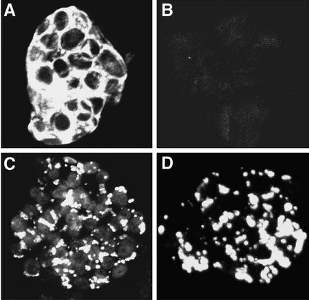
Figure 5.
Immunofluorescence localization of MerB and ER-MerB in fixed Arabidopsis leaf cells. A, MerB WT line. B, MerA line with no MerB (negative control). C and D, ER-MerB line. The confocal images shown are reconstructed from stacks of 0.5-micron photographs. Under equivalent laser parameters, MerB (A) and ER-MerB (C and D) showed different patterns of localization. The dimension of each cell is approximately 25 × 40 μm.

