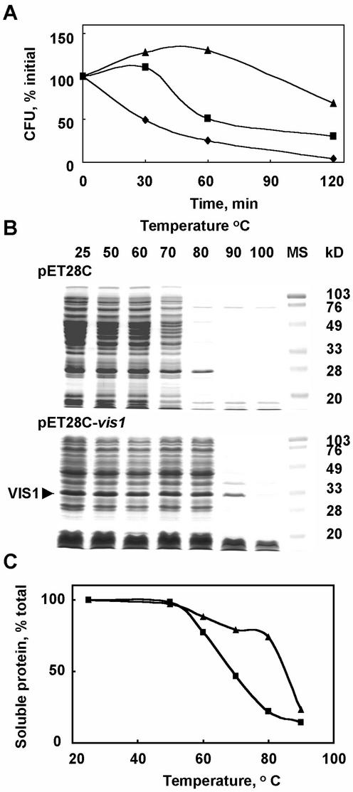
Figure 8.
Expression of vis1 in E. coli increases cell thermo-tolerance (A) and prevents aggregation of the bacterial protein at elevated temperatures (B and C). A, E. coli BL21 (DE3) harboring either the pET28C or pET28C-vis1 were grown at 27°C in Luria-Bertani (LB) plus 50 mg L−1 kanamycin to the initial OD600 of 0.333 (pET28c), 0.205 (pET28c-vis1, without isopropylthio-β-galactoside [IPTG]), and 0.147 (pET28c-vis1, 1 mm IPTG induced for 1 h) and shifted to 50°C. At the indicated time intervals, samples were withdrawn, and the viable cell count was determined using appropriate dilutions on LB plates containing kanamycin after incubation at 37°C overnight. Shown are the viable cell counts for E. coli BL21 (DE3) harboring the pET28C (♦), pET28C-vis1 in the absence of IPTG (▪), and presence of IPTG (▴). B, The bacterial cells harboring either the pET28C or pET28C-vis1 were grown at 27°C in LB medium plus 50 mg L−1 kanamycin in the presence of 1 mm IPTG for 1 h as described above and pelleted by centrifugation (12,000g) for 5 min. Cell pellets were resuspended in a buffer containing 25 mm Tris-HCl, pH 7.5, 10% (v/v) glycerol, 2 mm dithiothreitol, and 1 mm EDTA, sonicated, and centrifuged for 10 min at 12,000g in a microcentrifuge to obtain soluble proteins. Aliquots of soluble protein were incubated at the indicated temperatures for 20 min, and supernatant was collected after centrifugation. Equal volume of supernatants was separated on SDS-PAGE. Shown are the Coomassie R-250-stained gels. Arrow indicates the VIS1 present in the bacterial extracts. C, The percent protein remaining soluble after heat treatment in samples described in B. Symbols are the same as in A. The total soluble protein was determined by the dye-binding assay kit from Bio-Rad (Hercules, CA) using bovine serum albumin as standard.

