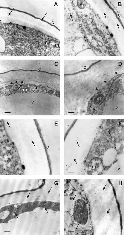
Figure 6.
PAO immunoelectron microscopy of epidermal cells from maize mesocotyls: effect of light exposure and auxin treatment. A and B, Portions of epidermal cells from etiolated mesocotyls showing scattered gold particles in the cytoplasm and the outer cell wall (arrows). C and D, Portions of epidermal cells from light-exposed maize mesocotyls with numerous gold particles in the cytoplasm and the outer cell wall. Note the preferential localization of labeling in the inner half of the cell wall and the presence of some grains in close proximity to the plasma membrane (arrowheads). In the cytoplasm, immunoparticles are sometimes found inside endoplasmic reticulum cisternae and vesicles (arrows). The cuticle, vacuole, and mitochondria are negative. E and F, Portions of epidermal cells from etiolated, NAA-treated maize mesocotyls. Few gold particles are found in the cytoplasm and the outer cell wall (arrows). The cuticle, vacuole, and mitochondria are unlabeled. G and H, Portions of epidermal cells from light-exposed, NAA-treated maize mesocotyl showing a moderate number of gold particles in the cytoplasm and the outer cell wall (arrows). Magnification: A, ×14,400, bar = 0.4 μm; B, ×21,600, bar = 0.25 μm; C, ×8,600, bar = 0.6 μm; D, ×21,600, bar = 0.25 μm; E, ×36,000, bar = 0.15 μm; F, ×36,000, bar = 0.15 μm; G, ×21,600, bar = 0.25 μm; H, ×21,600, bar = 0.25 μm. c, Cuticle; v, vacuole; m, mitochondrion. Micrographs shown are representative fields of sections obtained from three independent experiments.

