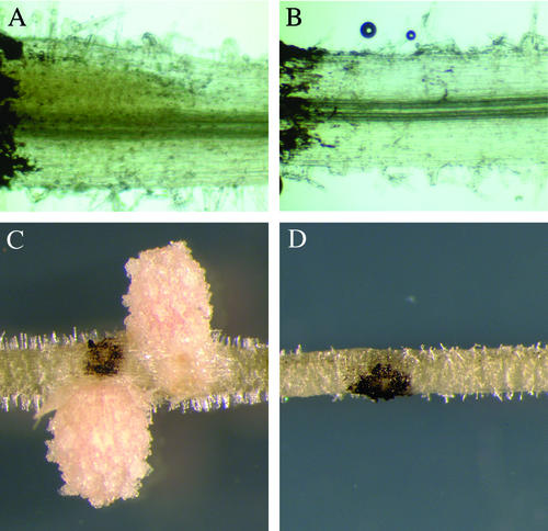
Figure 2.
The mitotic induction of cortical cells by S. meliloti is absent in nsp2. Plants were spot inoculated with S. meliloti and assessed at 3 d postinoculation (A and B) and 18 d postinoculation (C and D). At 3 d postinoculation, roots were stained with 0.1 m potassium iodide that causes a diffuse yellow staining within dividing cortical cells in wild-type plants (A) that is completely absent in nsp2-2 plants (B). C, Nodules form at later time points within the restricted area of the inoculation in wild type; D, no response is apparent at these later time points in nsp2. The dark coloration in all the images is lamp black that was applied to mark the point of inoculation.

