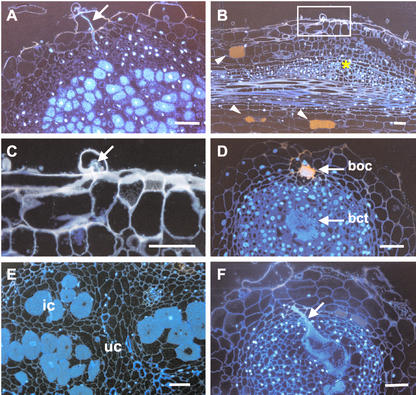
Figure 7.
Fluorescent micrographs of infection events in wild type and crinkle after 2 weeks (A–C) and 1 month (D–F) infection with M. loti. A, Developing nodule of wild type showing the infection thread and infected cells; B, bump of crinkle with arrested infection; C, close-up of thin section indicated in B; D, empty nodule (type I) of mutant showing aggregated bacteria in the intercellular spaces at the outer cortex (boc) and at the central tissue (bct); E, type II nodule of mutant containing infected cells scattered in the central tissue; F, Enlarged infection thread in the alb1-1 nodule. Arrows, Infection thread; asterisk, cortical cell division; arrowheads, pigmented cortical cells; ic, infected cells; uc, uninfected cells. Bars = 50 μm.

