Figure 4.
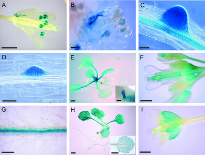
Histochemical localization of LRX expression in LRX promoter::uidA transgenic plants. A and B, pPEX3::uidA expression. C, pLRX6::uidA expression. D through F, pLRX5::uidA expression. G through I, pLRX4::uidA expression. A, Open flower. Mature pollen grains show a strong expression of pPEX3::uidA. B, Pollen germinating on a stigma. C, Emerging lateral roots, showing strong expression of pLRX6::uidA in the lateral root meristem, as well as in the vascular tissue of the mature primary root. D, Root of a 7-d-old seedling; GUS activity is restricted to the emerging lateral roots. E, Fourteen-day-old seedlings; pLRX5::uidA is expressed in the leaf primordia and stipules (inset image) at the basis of the rosette leaves. A moderate activity persisted during further expansion of the leaves, preferentially in petioles. F, Opened flowers and flower buds. GUS activity is detected in the carpels and faintly in the pedicels. Carpel expression seems higher in the style directly under the stigma and at the base of the organ and the junction with the receptacle (abscission zone). G, Primary mature roots; the vascular bundle is strongly stained. H, Fourteen-day-old seedlings; the inset picture shows a leaf stained for a shorter time to reveal the stronger expression in vascular tissue. I, Open flower; GUS activity is detected in sepals, mostly in the veins. Bars = 1 mm (A, F, and I), 50 μm (B–D), 2 mm (E and H), 350 μm (E, inset image), and 200 μm (G).

