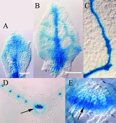
Figure 6.
AtHB20::GUS expression in leaves. A, First rosette leaf primordium at 3 DAG: diffuse expression with elevated levels along I1° and at the tip of the primordium. B, 4 DAG: strong expression along the differentiating midvein and weak expression along I2°s. C, Highly localized and strong expression at late stage of secondary vein differentiation. D, Cross section of a 7-DAG leaf primordium: Expression is confined to cells in vascular bundles. E, Higher magnification of midvein in D (arrow) shows strong expression in fascicular cambium (arrow in E).

