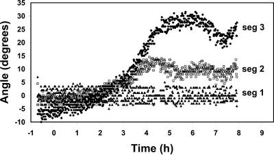
Figure 5.
Localized changes in root orientation following illumination with unilateral red light. The data show the angles of orientation of different regions of a representative root when the apical segment was constrained at 0° (vertical). The apical most portion of the root is segment 1, and each segment (seg) is 330 μm in length. Most of the photocurvature can be seen to result from curvature of segment 3. This experiment was repeated 12 times with similar results.

