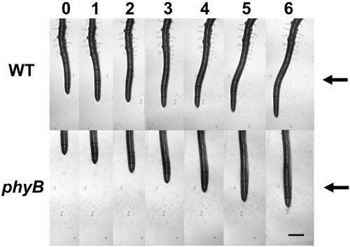
Figure 6.
Images of roots of WT and phyB seedlings following illumination with unilateral red light. In this experiment, red light was applied from the direction indicated (arrow) at time 0, and the root tip was constrained at 0° (vertical) by the feedback system. Images were taken hourly as indicated along the top of the figure. Note the obvious curvature in the WT root while the root of the phyB mutant grew straight, without a response to the red light. Scale bar = 500 μm.

