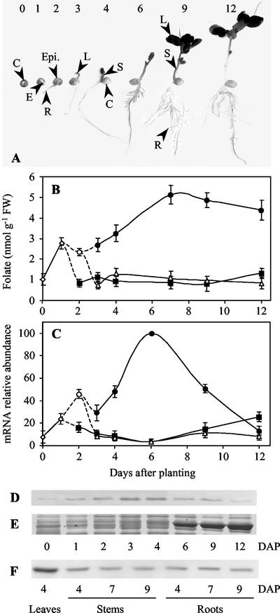
Figure 4.
Changes in folate content and HPPK-DHPS mRNA and protein expression in pea organs during development in the light. A, Pea seedlings at different stages of development. Imbibed seeds were planted in vermiculite at d 0 and grown at 22°C with a photoperiod of 12 h. At d 0 and 1, embryos (E) could be analyzed apart from the cotyledons (C). From d 2, the embryos could be separated into the epicotyls (Epi.) and the roots (R). The separation of stems (S) and leaves (L) occurred at d 3. The first leaves to appear are followed during all of the examined period of growth. B, Changes in folate content in pea organs during development. Folate was determined in embryos (⋄), epicotyls (○), roots (▪), leaves (●), and stems (▵) using a microbiological assay with L. casei. C, HPPK-DHPS mRNA expression in pea organs during development. The amount of HPPK-DHPS mRNA was estimated by semiquantitative RT-PCR using total RNA isolated from the different organs (symbols are similar to B). The amount of mRNA detected in 6-d-old leaves was the highest and was used as a reference for comparative analysis. D through F, HPPK-DHPS protein expression in pea organs during development. D, Thirty micrograms of soluble proteins extracted from embryos (d 0 and 1), epicotyls (d 2), and leaves (d 3–12) were analyzed by western blot with antiserum to HPPK-DHPS. E, Staining with Coomassie Brilliant Blue (polypeptides in the 50–60 kD range) illustrates the accumulation of the large subunit of Rubisco (about 55 kD) during leaf development. This polypeptide migrates just above HPPK-DHPS (52 kD), thus packing this protein when it accumulates. Therefore, from d 6, the amount of HPPK-DHPS is probably underestimated. F, Proteins (30 μg) from 4-d-old leaves, stems, and roots (d 4–9) are analyzed by western blotting with HPPK-DHPS antiserum. The data shown in this figure are means ± se from at least two determinations using three independent cultures of pea seedlings. DAP, Days after planting.

