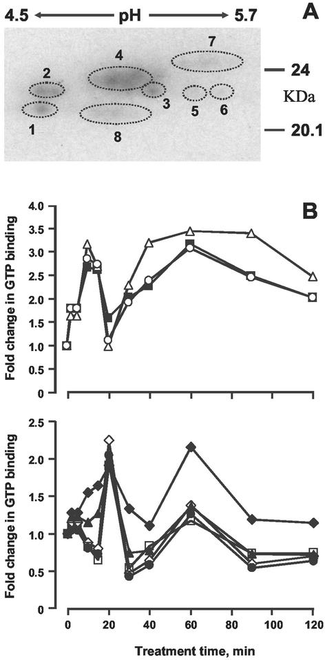
Figure 3.
Two-dimensional separations of G-proteins. Proteins were extracted with 1% (w/v) Triton X-100 from untreated or ethylene-treated (1 μL L−1) epicotyls and separated by two-dimensional gel electrophoresis. A, Designation of spots; B, quantification of GTP binding during time course: 1 (⋄), 2 (●), 3 (♦), 4 (▴), 5 (▪), 6 (▵), 7 (○), 8 (□).

